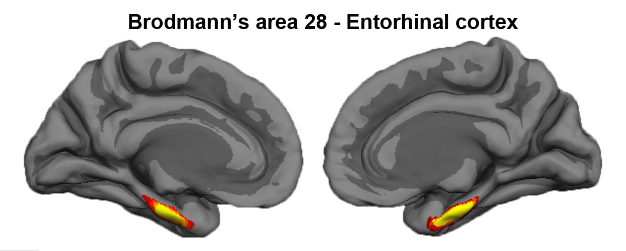
Entorhinal Cortex

The entorhinal cortex is located on the crown of the anterior parahippocampal gyrus. Brodmann assigned the number 28 to entorhinal cortex (Brodmann’s area 28). Entorhinal cortex displays a unique cytoarchitecture with large neurons in layer II that form clusters. The entorhinal cortex is the gateway to the hippocampus. In Alzheimer’s disease (AD), neurons in the entorhinal cortex, particularly those in layer II, begin to die and some leave behind a pathological marker – the neurofibrillary tangle. Neurofibrillary tangles and significant cell death characterize the neuronal loss in AD where neurofibrillary tangles and subsequent atrophy have been correlated with dementia.
Please note that there two entorhinal labels exist in FreeSurfer 1) the EC label included in the aparc atlas and 2) the cytoarchitecturally-defined entorhinal label (i.e., the ex vivo one).
Reference for Entorhinal cortex, Cytoarchitecturally-defined labels:
Fischl B, et al. Predicting the location of entorhinal cortex from MRI. Neuroimage. 2009 Aug 1;47(1):8-17.
Where to find the entorhinal (cytoarchitectural-defined) labels in FreeSurfer
Right and left labels for entorhinal are available in freesurfer 5.3
The labels are in freesurfer surface space and located in subjects dir subj/label/?h.entorhinal_exvivo.label
See instructions on how to view in freeview
To generate stats on this label (../stats/$hemi.BA.stats)
