|
Size: 23949
Comment:
|
← Revision 103 as of 2026-10-28 18:50:43 ⇥
Size: 21300
Comment:
|
| Deletions are marked like this. | Additions are marked like this. |
| Line 4: | Line 4: |
| == Citation == | |
| Line 5: | Line 6: |
| * [[https://doi.org/10.1101/2023.06.08.544050|Machine learning of dissection photographs and surface scanning for quantitative 3D neuropathology]]. H Gazula, H Tregidgo, B Billot, Y Balbastre, J Williams Ramirez, R Herisse, LJ Deden-Binder, A Casamitjana, EJ Melief, CS Latimer, MD Kilgore, M Montine, E Robinson, E Blackburn, MS Marshall, TR Connors, DH Oakley, MP Frosh, SI Young, K Van Leemput, AV Dalca, B Fischl, CL Mac Donald, CD Keene, B Hyman, and JE Iglesias. Under revision.) [[https://www.youtube.com/watch?v=wo5meYRaGUY|Click here for a video with an overview of the method]]<<BR>> {{attachment:video_screenshot.png||width="666",}} <<BR>><<BR>> |
* Gazula, H., Tregidgo, H.F., Billot, B., Balbastre, Y., Williams Ramirez, J., Herisse, R., Deden-Binder, L.J., Casamitjana, A., Melief, E.J., Latimer, C.S. and Kilgore, M.D., 2024. Machine learning of dissection photographs and surface scanning for quantitative 3D neuropathology. Elife, 12, p.RP91398. [[https://doi.org/10.7554/eLife.91398.4|doi]] The machine learning slice imputation in the reconstruction is described in: * Crespo-Aguirre, M., Williams-Ramirez, J., Zemlyanker, D., Hu, X., Deden-Binder, L.J., Herisse, R., Montine, M., Connors, T.R., Mount, C., !MacDonald, C.L., Keene, C.D., Latimer, C.S., Oakley, D.H., Hyman, B.T., Lawry-Aguila, A. and Iglesias, J.E., 2026. Improving neuropathological reconstruction fidelity via AI slice imputation. Acta Neuropathologica Communications. [[https://doi.org/10.1186/s40478-026-02451-3|doi]] Automatic segmentation model trained using nnUNet: * Williams Ramirez, J., Zemlyanker, D., Deden-Binder, L., Herisse, R., Garcia Pallares, E., Gopinath, K., Gazula, H., Mount, C., Kozanno, L.N., Marshall, M.S., Connors, T.R., Frosch, M.P., Montine, M., Oakley, D.H., Mac Donald, C.L., Keene, C.D., Hyman, B.T., Fischl, B. and Iglesias, J.E., 2026. A public generalizable AI tool for automated segmentation of coronal brain tissue slabs for 3D neuropathology. PLOS One, 21(8), p.e0355740. [[https://doi.org/10.1371/journal.pone.0355740|doi]] * Isensee, F., Jaeger, P.F., Kohl, S.A., Petersen, J. and Maier-Hein, K.H., 2021. nnU-Net: a self-configuring method for deep learning-based biomedical image segmentation. Nature methods, 18(2), pp.203-211. [[https://www.youtube.com/watch?v=wo5meYRaGUY|Click here for a video with an overview of the method]]<<BR>> {{attachment:video_screenshot.png||width="666"}} <<BR>><<BR>> |
| Line 13: | Line 21: |
| This manual provides step-by-step instructions for using a 3D scanner on ex-vivo brain specimens, preparing tissue slabs for gross photography, and outlining the data processing pipeline to create a 3D reconstruction of brain slab images using the acquired scan as a reference surface.<<BR>><<BR>> |
This manual provides an end-to-end guide for using the tools available in FreeSurfer to reconstruct dissection photographs of brain slabs into a 3D reconstruction for quantitative analysis. Reconstructions are done by registering photographs to a 3D reference such as an MRI, a post-mortem surface scan, or an atlas. The guide covers acquiring surface scans prior to slabbing, preparing tissue slabs for gross photography, and preprocessing photographs using the dissection_photo graphical user interface.<<BR>><<BR>> == Requirements == * The latest FreeSurfer development version sourced in your terminal. * Model files (downloaded once): * the [[https://ftp.nmr.mgh.harvard.edu/pub/dist/lcnpublic/dist/dissection_photo_model/photo_imputation_unet.pth|reconstruction model]], downloaded into `$FREESURFER_HOME/models` * A CUDA-enabled graphics card is optional: segmentation and reconstruction are much faster with one. == Overview == 1. '''Acquire a 3D reference''': either a surface scan or an MRI. Without one, slabs are registered to an atlas. 1. '''Photograph the tissue slabs''' 1. '''Preprocess the photos in the dissection photo GUI''' 1. '''Reconstruct the 3D volume''' 1. '''Downstream analysis of photographs''' = Part 1: Acquisition = |
| Line 16: | Line 38: |
| A surface scan of the specimen, taken before it is cut, is the most accurate reference for the reconstruction. The full scanning protocol (scanner setup, sample setup, handheld scanning, exposure, and editing and meshing the point clouds) is on its own page: '''[[PhotoTools Surface Scanning|Surface Scanning Protocol]]'''. What the reconstruction needs from the mesh: |
A surface scan of the specimen is taken after the cerebellum has been sectioned off and before the specimen is slabbed. The full scanning protocol is available on its own page: '''[[PhotoTools Surface Scanning|Surface Scanning Protocol]]'''. Summary of the key requirements of a reference mesh: |
| Line 21: | Line 44: |
| * A watertight mesh, saved as `.stl`. Small holes in deep sulci are fine. |
* A watertight mesh, saved as `.stl`. Small holes in the initial point cloud due to deep sulci are fine; these will be covered by the meshing process. |
| Line 26: | Line 48: |
| == Sample Setup == Post sectioning of the cerebellum, we obtain a surface scan of the specimen before then sectioning it into slabs for photography. We place a glass sheet atop to protect the turntable and ease cleanup. The glass sheet contains markers that be used for tracking. The specimen is placed in the middle of the turntable so that the scanner gets the best view of the sample as it rotates. Occluding the turntable markers will also affect the stitching of scans if the scan is set to “Turntable Coded Targets.” When using custom markers however, which we recommend, the turntable targets should be covered completely. When using custom markers in either fixed or handheld mode, make sure the specimen is not covering any markers on the glass sheet. {{attachment:05_SamplePlacement.png||width="435",height="445"}} <<BR>>''Sample placement on the turntable with sample lateral side down.''<<BR>> Two scan projects are created for all specimens at opposite orientations. For hemispheres, we first acquire a scan with the sample medial side down. Next, the hemisphere is flipped to be lateral side down. Again, to reduce sample movement, we wipe down excess fixative on the sheet of glass to improve traction. Whole Brains are scanned upright (brain stem down) and upside down. Although these types of samples do not tend to move during the scan, they can deform between the two orientations, which can lead to small errors in the final surface reference. '''''Note''''': Samples should have the cerebellum removed before acquisition. The cerebellum is not considered in the reconstruction as it is not on the imaged slabs, including it in the scan will lead to incorrect slice reconstruction. {{attachment:06_ScanIncorrect.png||width="435",height="380"}} <<BR>>''Scan incorrectly including the cerebellum.''<<BR>> <<BR>> == Handheld Scanning == Handheld mode can be particularly useful for fresh tissue, since it cannot maintain its shape or be flipped for two-sided scans because of deformation. In this mode, a transparent surface can be used to scan both sides of the sample without having to shift the easily deformable tissue. {{attachment:07_ExampleHandheld.png||width="435",height="404"}} <<BR>>''Example of a handheld scan setup.''<<BR>> == Running the Scan == In the scan workspace, use the preview window to center the tissue in the center of the field of view. Set the red crosshairs to the middle of the specimen for full coverage as the turntable rotates. {{attachment:10_expsure_good.png||width="288",height="250"}} <<BR>>''Scanner preview.''<<BR>> The camera exposure has a large effect on scan quality. Incorrect exposure can lead to noisier surfaces in the resulting scan point clouds. Adjust exposure in the main scan menu under the camera preview screen. {{attachment:09_ScannerExp.png||height="72"}} <<BR>>''Scanner exposure slider.''<<BR>> Use the preview window to check exposure, areas that are overexposed will appear red. A good exposure will show some red areas but too many, see below for a reference of a good exposure. For specimens with particularly difficult lighting HDR mode can be used to better render darker features. || {{attachment:11_exposure_over.png||width="245",height="221"}} || {{attachment:12_exposure_under.png||width="237",height="215"}} || {{attachment:10_expsure_good.png||width="246",height="212"}} || ||''Overexposed (red filter on sample + bright image)'' ||''Underexposed (black filter on sample + dark image)'' ||''Sufficient exposure (can see sample, markers, decent contrast)'' ||<<BR>> === HDR Mode === HDR mode is used for samples with dark features like bruising or veins. This mode captures three images at each turntable step at different exposures which better capture darker and deeper structures in the tissue (e.g., sometimes this mode is good if point clouds aren’t generated near medial structures, including 3^rd^ventricle, septum pellucidum, corpus callosum, parahippocampal gyrus, occipitotemporal gyrus, and any optic structures – nerve, chiasm) {{attachment:13_dark_veins.png||width="432",height="463"}} <<BR>>''Sample with dark veins.''<<BR>> == Editing and Saving Point Clouds == === Removing Excess Elements === 1. Remove excess data or turntable markers using the deleting tool if the scanner picks up other neighboring geometry. 1. Select extra elements by holding the shift key and left click. 1. Delete these points by clicking the “Delete Selected Data” button or pressing the delete key. 1. Press the “Complete Editing” check-mark icon once all necessary edits are done. 1. Note: Editing point clouds is easier than editing meshes so make sure to remove any excess elements before generating a mesh. {{attachment:14_UI_guide.png||width="667",height="394"}} <<BR>>''Performing the second run after removing excess elements from the first set of point clouds.''<<BR>> 1. When finished editing, a point cloud will be generated, which should have saved automatically as “Project 1.” 1. Briefly QA scan data, looking at noise or holes in the surface of the scan. Some gaps in the point clouds are normal. Any scan can be redone or added onto to get better results at any time. <<BR>> === Aligning both runs === If you have two scanning sessions, there are a couple of ways the point-clouds can be registered, an automatic and manual method. Only use manual alignment if the automatic method fails. <<BR>> 1. Click the alignment icon and select both scanning sessions. Click the automatic alignment button at the top left of the screen. If the alignment fails, continue with the next steps. 1. Hold the shift key and left click to select three collinear points on each scan session. Ideally, we would want to place one point on the anterior, superior, and posterior sides of the brain. Spread out points as much as possible to get the best alignment. 1. Click Complete when finished with the alignment. <<BR>> === Creating a Mesh === 1. Click on the global optimization and confirm the resulting point cloud. 1. Click on the mesh icon to generate a mesh model from your generated point clouds. 1. Select watertight, which will create a mesh that is closed and has no holes. 1. Choose the “high-detail” option for the best mesh resolution. 1. Select "No filter" so no smoothing is applied on the mesh. 1. This reconstruction should take approximately 3+ minutes, depending on the equipment. 1. Save your mesh when reconstruction has finished. Your mesh should be saved as a .stl file automatically, but it is also recommended to save your point clouds (.asc files). <<BR>> === Addressing mesh quality === When acquiring the mesh, the sides of the sample are critical for proper point cloud alignment. Poorly captured sides will lead to inaccurate alignments between scans, which will cause issues in the final reconstruction. The following example shows a handheld scan where the sides of the tissue were not acquired, resulting in a flatter/thinner appearance which will negatively affect the quality of the final reconstruction. {{attachment:15_bad_mesh.png||width="436",height="330"}} <<BR>>''Mesh missing sides of the top scan.''<<BR>> Ideally, the sides of the mesh should be captured as shown in the following example. When performing a handheld scan, it can be difficult to acquire the sides of the sample without acquiring other surfaces. These additional surfaces can be edited and deleted in the same way markers are removed. {{attachment:16_good_mesh.png||width="438",height="329"}} <<BR>>''Mesh correctly including the sides of the tissue.''<<BR>> Note that the previous surface has some holes due to deep sulci, which should not pose a problem with the reconstruction. Watertight meshing should be able to handle small gaps in the point-cloud. <<BR>><<BR>> = Preparing Slab Data = |
== Preparing Slabs == |
| Line 110: | Line 54: |
| Use a high-contrast background that stands out in uniform contrast to the brain slabs, such as a solid black background. This will make slabs are easier to segment downstream. Using a complex background with color similar to your brain slabs, will lead to a more manual intervention such as having to manually trace contours, which is not only time-consuming but also non-reproducible.<<BR>> |
Use a high-contrast background that stands out in uniform contrast to the brain slabs, such as a solid black background. This will make the slabs easier to segment downstream. Using a complex background with a color similar to your brain slabs will lead to more manual intervention, such as having to manually trace contours, which is not only time-consuming but also non-reproducible.<<BR>> |
| Line 113: | Line 56: |
| ||Difficult to segment low contrast background. ||Easy to segment high contrast background. || Additionally, all tissue block faces must be facing the same direction. All slices should be either from an anterior or a posterior view. Note if slabs are from the posterior view, an extra flag, '''--photos_of_posterior_side''' must be used for the final reconstruction. When a single slice produces multiple tissue blocks, they should be placed as similar to the original anatomy as possible. For example, after acquiring slices from the frontal pole, the initial blocks corresponding to the temporal lobe will often be detached from the main block. The following figure shows a) Proper anatomical placement of tissue and, b) Tissue placed too far apart, which is not anatomically accurate. {{attachment:19_slab_placement.png||width="667",height="483"}} <<BR>>''Tissue slab placement a) Properly placed tissue b) Tissue placed in a non-anatomical manner.''<<BR>> Another example of difficult tissue placement can be the corpus callosum. In the following figure, we see a good example setup with high contrast for the background and good lighting. However, the placement of the corpus callosum on the rightmost slab is incorrect, which will negatively affect volumetric estimates of the third ventricle. {{attachment:20_incorrect_tissue_placement.png||width="659",height="333"}} <<BR>> <<BR>> |
||Low-contrast background: difficult to segment. ||High-contrast background: easy to segment. || Additionally, all tissue block faces must face the same direction: all slices should be photographed either from the anterior or from the posterior view. Note that if slabs are photographed from the posterior view, the extra flag '''--photos_of_posterior_side''' must be used in the final reconstruction command. When a single slice produces multiple tissue blocks, they should be placed as close to the original anatomy as possible. For example, after acquiring slices from the frontal pole, the initial blocks corresponding to the temporal lobe will often be detached from the main block. The following figure shows a) proper anatomical placement of tissue, and b) tissue placed too far apart, which is not anatomically accurate. {{attachment:19_slab_placement.png||height="483",width="667"}} <<BR>>''Tissue slab placement: a) properly placed tissue; b) tissue placed in a non-anatomical manner.''<<BR>> Another example of difficult tissue placement is the corpus callosum. The following figure shows a good setup, with a high-contrast background and good lighting. However, the placement of the corpus callosum on the rightmost slab is incorrect, which will negatively affect volumetric estimates of the third ventricle. {{attachment:20_incorrect_tissue_placement.png||height="333",width="659"}} <<BR>> <<BR>> |
| Line 128: | Line 69: |
| To calibrate pixel size, we will need landmarks with known distances in between them to be provided by the user. The minimum requirement is a ruler, which provides us with two discrete landmarks. This enables rough This method is preferable for legacy images that only have a ruler for reference. {{attachment:21_twoPoint.jpeg||width="664",height="382"}} <<BR>> '''''Tip''''': It is better to use landmarks that are as far apart as possible, as the relative error when manually setting the reference in pixels per millimeter decreases with greater distances. A more effective setup involves four landmarks placed the corners of a rectangle of known dimensions. This enables us to correct for both pixel size and perspective. In the example below, we can easily tell that the 4 points marked on the grid define a rectangle of dimensions 170x200 mm. {{attachment:22_fourPoint.png||width="659",height="561"}} <<BR>> The optimal scenario is to have 4 points defined by fiducial markers with highly salient features. This enables our software to automatically recognize the fiducials, which minimizes human interaction, reducing time and maximizing reproducibility. We '''strongly''' recommend that you print these set fiducials and place them on the corners of the board. {{attachment:23_fiducials.png||width="416",height="439"}} <<BR>> '''''Tip''''': For best results, affix these fiducials to a small piece of rubber/wood/plastic that lifts them X mm above the board, where X is the thickness of your typical brain slice. Lifting the fiducial minimizes error due to the surface of the brain slice not being at the same distance as the plane of the board surface, but rather a few millimeters above it. {{attachment:24_fiducialTissue.png||width="674",height="460"}} <<BR>> The fiducial form the four corners of our corrected image; hence all tissue should be within the rectangle created from the four fiducial corners. The following image shows slab photos where the fiducials were not placed sufficiently apart. {{attachment:25_tissueSpace.png||width="685",height="404"}} <<BR>> By drawing a rectangle with all fiducial markers, it is clear that much of the tissue lies outside of the pixel-corrected region. {{attachment:26_tissueBox.png||width="683",height="403"}} <<BR>> In summary, here is an example of what an optimal photographic setup would look like, including the uniform, high-contrast background and provided fiducials located at known distances, and consistent lighting on all slabs. {{attachment:27_goodContrast.png||width="676",height="478"}} <<BR>> '''''Tip''''': It does not matter if slabs are not presented on a grid, as long as the distance between the fiducials is known. The software will draw digital rulers for your images later pixel-correction.<<BR>><<BR>> = Recommended Directory Structure = We recommend the following directory structure for data and case organization. Each case should have its own directory, and within each case directory, create the following seven subdirectories to store the respective outputs: 1. photos – Brain slab images (.JPG, .PNG). 1. deformed - Pixel-corrected images. 1. point_clouds – Files from the project collected from the surface scanner. 1. mesh – Watertight mesh processed from point clouds on the scanner (.stl). 1. masked – Binarized masks created from pixel-corrected photos. 1. connected_components - .npz files showing the connected components for each image. 1. recon - Final reconstructed volumes, and segmentations. Note that during the image preprocessing GUI files under deformed, masked, and connected_components will be automatically generated. After each step, ensure that your data is saved to the appropriate subdirectory. Below is an example of how files should be organized in the recommended structure: <<BR>> || {{attachment:28_demoDirectory.png||height="555"}} || {{attachment:29_demoDirectory.png||height="180"}} <<BR>> {{attachment:29.2_demoDirectory.png||height="203"}} || <<BR>><<BR>> = Photo Preprocessing = The following guide will walk through the steps to preprocessing the brain slab images. This includes pixel correction, binarizing images, and selecting connected components. Make sure Freesurfer is properly sourced, and you have downloaded the model weights before continuing with the tutorial. <<BR>> == Pixel Correction Using Fiducials == Using the provided fiducial markers, we can correct for non-uniformities in pixel size across the images. If not available this step can be skipped retrospective correction modes can be used in the '''dissection_photo_gui''' covered in the next section. For images with fiducials, a calibration is required for pixel correction. For best results, use an image of the setup without tissue.<<BR>> To start the fiducial calibration use command '''fiducials_calibration''' {{attachment:30_fiducialCalibIntro.png||width="550",height="275"}} <<BR>> After loading the image, start the calibration process. {{attachment:31_fiducialCalibWindow.png||width="548",height="440"}} <<BR>> Pan and zoom can be used to have a precise selection in these GUIs. First, select the center of one of the fiducials, followed by selecting the outer ring. || {{attachment:32_fiducialCenter.png||height="237"}} || {{attachment:33_fiducialDiameter.png||height="237"}} || ||The first selection at the fiducial center. ||The second selection at the outer circle of the fiducial. || After selecting all the points, set the width and height between the fiducial markers. Next, click “Perform calibration,” to save a calibration '.npz' file. This calibration file will be used for automating pixel correction in subsequent steps, as long as the fiducial markers are not moved. <<BR>> == Dissection Photo GUI == The dissection photo GUI performs all preprocessing tasks, including pixel correction, binarizing images, and selecting connected components. To run the GUI, source Freesurfer and use command '''dissection_photo_gui'''. The first time the GUI is run it will display instructions on setup of model weights for the segmentation step.<<BR>> == Pixel Correction == In the first menu of the '''dissection_photo_gui''' select the input directory with the unprocessed gross photographs. Photographs should be set in the order such that tissue slabs are seen in the order they were cut. Additionally select the output directory where the pixel-corrected images will be saved. Here we have the option to load an optimal calibration file if available to automate pixel correction. Select '''Go!''' when you have defined the required directories. <<BR>> {{attachment:35_dissection_photo_gui_screen_1.png||width="600",height="400"}} <<BR>> If a calibration file was used the next widow will display the pixel-corrected images for quality assurance purposes. Click '''Next''' to proceed assuring the images were corrected.<<BR>><<BR>> If no calibration file is provided the next window will be for the '''Retrospective Correction''' mode. Select the type of calibration, 2-point, 3-point, or 4-point, and its relevant dimensions in millimeters. The 2-point calibration is used when there is a single axis of ground truth measurement, which enables rough pixel size correction (e.g., one ruler in the image). The 3-point calibration is used when there are three points on the corners of a rectangle – but the fourth point is not available (e.g., there are two orthogonal rulers). The 4-point version should be chosen if the four corners of a rectangle can be identified; this is the default model when the automatic mode with calibration file is used. The 3- and 4-point versions enable perspective correction (approximate with 3 points; exact with 4). We can pan and zoom to make precise selections on the image.<<BR>> || {{attachment:36_dissection_photo_gui_screen_2.png||width="400",height="266"}} || {{attachment:37_dissection_photo_gui_screen_2_zoomed.png||width="400",height="266"}} || |
To calibrate pixel size, the user needs to provide landmarks with known distances between them. The minimum requirement is a ruler, which provides two discrete landmarks. This enables rough pixel size correction. This method is preferable for legacy images that only have a ruler for reference. {{attachment:21_twoPoint.jpeg||height="382",width="664"}} <<BR>> '''''Tip''''': It is better to use landmarks that are as far apart as possible, as the relative error when manually setting the reference in pixels per millimeter decreases with greater distances. A more effective setup uses four landmarks placed at the corners of a rectangle of known dimensions. This enables us to correct for both pixel size and perspective. In the example below, we can easily tell that the 4 points marked on the grid define a rectangle of 170 x 200 mm. {{attachment:22_fourPoint.png||height="561",width="659"}} <<BR>> The optimal scenario is to have 4 points defined by fiducial markers with highly salient features. This enables our software to recognize the fiducials automatically, which minimizes human interaction, reducing time and maximizing reproducibility. We '''strongly''' recommend that you print this set of fiducials and place them at the corners of the board. {{attachment:23_fiducials.png||height="439",width="416"}} <<BR>> '''''Tip''''': For best results, affix the fiducials to a small piece of rubber, wood or plastic that lifts them X mm above the board, where X is the thickness of your typical brain slice. Lifting the fiducials minimizes the error caused by the surface of the brain slice not being in the same plane as the board, but a few millimeters above it. {{attachment:24_fiducialTissue.png||height="460",width="674"}} <<BR>> The fiducials form the four corners of the corrected image; hence, all tissue should be within the rectangle formed by the four fiducials. The following image shows slab photos where the fiducials were not placed far enough apart. {{attachment:25_tissueSpace.png||height="404",width="685"}} <<BR>> Drawing a rectangle through the four fiducial markers makes it clear that much of the tissue lies outside of the pixel-corrected region. {{attachment:26_tissueBox.png||height="403",width="683"}} <<BR>> In summary, here is an example of an optimal photographic setup: a uniform, high-contrast background, the provided fiducials located at known distances, and consistent lighting on all slabs. {{attachment:27_goodContrast.png||height="478",width="676"}} <<BR>> '''''Tip''''': It does not matter if the slabs are not placed on a grid, as long as the distance between the fiducials is known. The software will draw digital rulers on your images during pixel correction.<<BR>><<BR>> = Part 2: Preprocessing Photographs with the Dissection Photo App = The dissection photo app has five principal windows for each subject: '''Add photos''', '''Correction''', '''Segmentation''', '''Connected components''' and '''Reconstruction'''. The step bar at the top of the window shows which step is being done. Users can backtrack by clicking any of the previous steps at any time, or click the house icon to return to the Projects screen with a summary of all reconstructions done or in process. For each project, work is saved as you go, so the app can be closed at any time and you can pick up later where you left off. Before continuing, make sure the development version of FreeSurfer is properly sourced, you have downloaded the model weights, and have set '''NNUNET_MODEL_DIR''' as an environment variable pointing to the segmentation model. To start the application, use the command '''dissection_photo_2'''. <<BR>> == Projects Page == Each specimen corresponds to a '''project''': a folder which will be populated with the outputs from all steps. The project home contains a summary of all projects and their progress. {{attachment:ds2_00_projects.png||width="560"}} <<BR>> ''Projects Home screen.'' * To start processing a new batch of dissection photographs click on '''New Project'''. * Open a project to continue where you left off or backtrack to any completed steps. '''Tip:''' For advanced users, the gear icon can be used to change which resources are used by the automatic segmentation model, covered in [[#Automatic_Segmentation_Settings|automatic segmentation settings]]. After naming a project you will be prompted to save a folder of the same name. It is in this folder that the outputs of all steps will be stored. == Directory Structure == For each project the following directory structure will be populated with all outputs. ||Folder / file ||Contents || ||`photos/` ||Copy of the original photographs || ||`deformed/` ||Pixel-corrected photos || ||`mask/` ||Binary tissue masks || ||`connected_components/` ||Label maps per slab || ||`recon/` ||The 3D-photo reconstruction || == Step 1. Add photographs == 1. Drag and drop a folder containing dissection photographs, or click '''Upload Images'''. 1. Select the photographs that should be used for the reconstruction. This can be used to omit repeat photographs, dissection photographs of cerebellar tissue, or gross photographs done prior to slabbing. Double click on any image in the grid for a larger preview. 1. If you have or would like to create a calibration file to automate pixel correction, click on '''+ Add a calibration file (optional)''' and follow the steps in [[#Optional:_Making_a_calibration_file|Making a Calibration File]] and then [[#Prospective_Correction_.28Using_fiducial_markers_and_calibration_file.29|Prospective Correction]]. 1. Click '''Get started'''. ||<:> {{attachment:ds2_01_add_photos_empty.png||width="420"}} <<BR>> ''Add photos, before any photographs are chosen.'' ||<:> {{attachment:ds2_02_add_photos_populated.png||width="420"}} <<BR>> ''Photographs added, with the calibration file box expanded.'' || === Optional: Making a calibration file === This step only needs to be done once, as long as the markers are not moved. For more information on proper usage refer to the [[#Markers_for_pixel_and_perspective_corrections|Markers for pixel and perspective corrections]] section in this wiki page. 1. On the Add photos menu, click '''+ Add a calibration file (optional)''' to expand the calibration file box, and click on '''Create one''' under the Upload Calibration button. 1. On the next menu, if you have not printed out the fiducial markers, they can be downloaded for printing by clicking the '''Download Marker Printout''' button. 1. Next, place markers at a known distance in a rectangle formation on the photography table. Be sure that opposite sides of the rectangle created by the fiducial markers are parallel; inaccurate marker placement can lead to incorrect perspective correction. 1. Upload the photograph of the empty dissection table with just the markers present, and enter the width and height of the fiducial box. 1. For each of the markers, click its centre and outer circle. Options to zoom, pan, and redo/undo can help fix mistakes and make the best calibration possible. 1. Click '''Save Calibration''' and save your calibration file in an easy-to-access location. '''Tip:''' Because of slight lens distortion, the overlaid labels for the outer circle of the markers might not always align throughout the whole circumference. This is not an issue. {{attachment:ds2_03_calibration_photo.png||width="640"}} <<BR>> ''The photograph of the empty table with the four markers, uploaded.'' ||<:> {{attachment:ds2_04_calibration_zoomed.png||width="420"}} <<BR>> ''Zoomed in on one marker, before clicking.'' ||<:> {{attachment:ds2_05_calibration_clicks.png||width="420"}} <<BR>> ''The marker's centre and outer circle clicked.'' || == Step 2: Pixel Correction == This step corrects all photographs to be at the same pixel size. Additionally, it can correct for perspective when using a calibration file, or when doing manual correction in 4-point mode. === Prospective Correction (Using fiducial markers and calibration file) === * Every photo is corrected automatically as soon as Correction opens. The markers are found in each photo separately, so it does not matter if the camera moved between photos. * Check each photo: '''Original''' shows the photo as taken, with the marker centres that were found; '''Corrected''' shows the result. A green tick in the strip at the bottom marks each corrected photo. * If the markers cannot be found in a photo, the app says which ones. On each of them, click the centres of the four markers on the Original view and click '''Correct this photo'''. === Retrospective Correction (Using a spatial reference within the photographs) === Choose how many points you can mark, enter the real distances, and place the points on each photo: ||'''Mode'''||'''You need'''||'''Corrects'''|| ||2 Point||One ruler or other known distance (entered as '''Ruler width (mm)''')||Pixel size only|| ||3 Point||Three corners of a rectangle, e.g. two rulers at right angles||Pixel size, approximate perspective|| ||4 Point||Four corners of a rectangle||Pixel size and perspective|| * Ctrl + left click places a point; once all are placed, Ctrl + click moves the nearest one. '''Clear points''' starts again. * Click '''Correct this photo''' (Ctrl + W also moves on to the next photo). Place points on the Original view; the Corrected view is for checking. ||<:> {{attachment:ds2_06_correction_ruler.png||width="420"}} <<BR>> ''2 Point mode: two points placed along a 150 mm ruler (Original).'' ||<:> {{attachment:ds2_07_correction_done.png||width="420"}} <<BR>> ''The corrected photo, with rulers (Corrected).'' || When every photo is corrected, make sure to click '''Continue to Segmentation'''. == Step 3: Binary Segmentation == While you were correcting, the app was already finding the tissue in each corrected photo. Here you check each mask and fix it. ||<:> {{attachment:ds2_08_segmentation_mask_on.png||width="440"}} <<BR>> ''The automatic mask (green) shown over the slabs.'' ||<:> {{attachment:ds2_09_segmentation_mask_off.png||width="346"}} <<BR>> ''The mask hidden ('''Show mask''' off); the note at the top lists the masks not yet previewed.'' || * '''Brush''' (Q) adds to the mask, '''Eraser''' (W) removes from it, and '''Fill''' (E) fills an area. Apply the tool with Ctrl + left drag; set the sizes with the sliders. * '''Mask opacity''' changes how see-through the mask is; '''Show mask''' (Ctrl + T) hides and shows it. Undo and redo with Ctrl + Z and Ctrl + Shift + Z. * Leave out everything that is not slab tissue: the background, labels, and fragments that do not belong to a slab. * '''IMPORTANT''': the cortical surface should not be part of the masks, so the first slab (if photographed from the anterior side) or the last slab (if photographed from the posterior side) should '''NOT''' be included. * You need to look at every photo's mask before '''Continue to Connected Components''' becomes available; a note at the top says which ones are left. === Automatic Segmentation Settings === Open them from the gear on the Projects screen. Changes apply the next time photos are segmented. {{attachment:ds2_10_seg_settings.png||width="480"}} <<BR>> ''Automatic segmentation settings.'' ||'''Setting'''||'''What it does'''|| ||Automatic segmentation (On / Off)||Off: every mask is drawn by hand from scratch. Only turn it off if the automatic segmentation fails on your photos.|| ||Model folds||Automatic: 5 folds on a GPU, 1 on the CPU. Five folds are steadier but 4-5x slower.|| ||Run on||Automatic uses the GPU if there is one; or choose GPU or CPU.|| ||Maximum threads||The most processor threads segmentation may use. If the computer has fewer, it uses all it has.|| ||Model folder||The folder of the segmentation model (`nnUNetTrainer__nnUNetPlans__2d`).|| == Step 4: Slab Connected Components == Here you tell the app which pieces of tissue belong to which slab. * In each photo, draw a box inside each slab (Ctrl + left drag), always in the same order: anterior to posterior, or posterior to anterior. The slab is colored as soon as its box is drawn. * Keep boxes small and inside the tissue, and do not let them overlap. If a slab is in several pieces (e.g. a detached temporal lobe), make sure its box touches every piece. * Click '''Save components''' (Ctrl + W also moves on to the next photo); moving to another photo saves it too. A green tick marks saved photos. If you change a saved photo, its tick goes away until you save it again. * Undo and redo with Ctrl + Z and Ctrl + Shift + Z. '''Slab overlay''' changes how strongly the slabs are colored. {{attachment:ds2_11_components_empty.png||width="420"}} <<BR>> ''Before any boxes are drawn.'' ||<:> {{attachment:ds2_12_components_good.png||width="420"}} <<BR>> ''One small box inside each slab; each slab is colored and the photo is saved.'' ||<:> {{attachment:ds2_13_components_bad.png||width="420"}} <<BR>> ''How '''NOT''' to draw boxes: they overlap each other and cover background.'' || == Step 5: Photo Reconstruction == Please see our separate page on the command [[mri_3d_photo_recon]]. The last step runs [[mri_3d_photo_recon]] on the images of the project. While the reconstruction is running in the background the user can continue processing images of other cases. 1. '''Output folder''': the folder inside the project for the results (default `recon`). 1. '''Slabs''': the hemisphere, the slab thickness in mm, which face of the slabs was photographed (anterior or posterior), the order the photos were taken in, and whether the tissue is fixed or fresh. 1. '''Reference''': none (the MNI atlas), a surface mesh (`.stl`), an MRI scan, or both. See [[#Which_reference_should_I_use.3F|Which reference should I use?]] 1. Optionally: the number of threads, a graphics card, and the advanced settings. Empty expert weights use the command's own defaults, shown in grey. 1. Click '''Run Reconstruction'''. The command being run is shown on the right ('''Copy command''' copies it, for example to run it on another computer), and its output appears in the log below it. The reconstruction keeps running if you switch to another project; the Projects screen shows its progress. {{attachment:ds2_14_reconstruction.png||width="640"}} <<BR>> ''A reconstruction running, with the command and its log.'' |
| Line 211: | Line 221: |
| After pixel correcting all images, click '''Proceed to Segmentation''' to continue to the next step.<<BR>> == Tissue Segmentation == After selecting an output directory for the masked images, our automatic segmentation tool will immediately start predicting a binary mask per each photograph. If a GPU is available all five folds of the model will be run which will slightly increase segmentation accuracy. After the first photograph is segmented by the model, the user can start cleaning the predicted binary masks. Here the user can pan and zoom like before, but can additionally use a brush tool to remove or add any pixels. Remove any non-slice tissue and the background. The opacity of the predicted mask can also be changed using the provided slider, or toggled using Ctrl+Z. <<BR>> || {{attachment:38_dissection_photo_gui_screen_3_noseg.png||width="400",height="266"}} || {{attachment:39_dissection_photo_gui_screen_3_seg.png||width="400",height="266"}} || <<BR>> '''IMPORTANT''': The cortical surface should not be part of the masks, so the first (if photographed from the anterior side) or last slice (if photographed from the posterior side) should '''NOT''' be considered. You can either leave it out of the mask or ignore it when processing with the GUI. <<BR>> Once we are satisfied with the mask, click '''Next''' to proceed to the next image. After all images have been segmented and cleaned, click '''Proceed to CC''' to continue to the connected components step. <<BR>> == Connected Components == Like previous steps, select an output directory for the connected components masks. The connected components section of the GUI will display the masked and pixel-corrected photographs. Here the user will draw boxes around the tissue slabs in each photograph. The GUI will then save the connected components as .npy files. <<BR>> {{attachment:40_dissection_photo_gui_screen_4_tissue.png||width="600",height="400"}} <<BR>> For each image select slices in order, either anterior-posterior or posterior-anterior. Remain consistent of slice order on all photographs. Connected components are selected by clicking and dragging to draw a box in a region of each individual slab in the photograph. After a box has been drawn in each individual slab, click '''Next ''' to save the output .npy file and move to the next image. '''Tip''': Make sure no boxes are overlapping. Once a box is drawn over the tissue, the slab will be colored to show what is being included as one slice.<<BR>> {{attachment:41_dissection_photo_gui_screen_4_color.png||width="600",height="400"}} <<BR>> Above is an example of how to correctly draw the boxes within the brain slabs themselves. These boxes inform the GUI that the pixels connected to these slabs are all part of the same tissue slice. Note the box covers tissue from the temporal lobe as it can sometimes be disjoint from the rest of the slab. <<BR>> Below is an example of how '''__NOT__''' to draw boxes for this GUI. Notice the occurrence of overlap between boxes and the inclusion of an abundance of black space. Try to keep the boxes drawn to small regions within the brain slabs themselves as to not include any background pixels. <<BR>> {{attachment:42_dissection_photo_gui_screen_4_bad.png||width="600",height="400"}} <<BR>><<BR>> = Photo Reconstruction = Please see our separate page on the command [[mri_3d_photo_recon]] <<BR>> = Photo-Synthseg Segmentation = |
= Part 3: Downstream Tasks = == Photo-Synthseg Segmentation == |
| Line 251: | Line 226: |
| '''Example for running SynthSeg on a Left Hemisphere''' <<BR>> . mri_synthseg \ . --i ./recon/photo_recon.mgz \ . --o ./recon/photo_recon_synthseg.mgz \ . --photo left \ . --parc |
'''Example for running SynthSeg on a Left Hemisphere''' <<BR>> . mri_synthseg \ ''''' ''''' . --i ./recon/photo_recon.mgz \ ''''' ''''' . --o ./recon/photo_recon_synthseg.mgz \ ''''' ''''' . --photo left \ ''''' ''''' . --parc ''''' ''''' |
Photo Reconstruction Documentation
Contents
- Photo Reconstruction Documentation
- Part 1: Acquisition
- Part 2: Preprocessing Photographs with the Dissection Photo App
- Part 3: Downstream Tasks
Citation
If you use these tools in your analysis, please cite:
Gazula, H., Tregidgo, H.F., Billot, B., Balbastre, Y., Williams Ramirez, J., Herisse, R., Deden-Binder, L.J., Casamitjana, A., Melief, E.J., Latimer, C.S. and Kilgore, M.D., 2024. Machine learning of dissection photographs and surface scanning for quantitative 3D neuropathology. Elife, 12, p.RP91398. doi
The machine learning slice imputation in the reconstruction is described in:
Crespo-Aguirre, M., Williams-Ramirez, J., Zemlyanker, D., Hu, X., Deden-Binder, L.J., Herisse, R., Montine, M., Connors, T.R., Mount, C., MacDonald, C.L., Keene, C.D., Latimer, C.S., Oakley, D.H., Hyman, B.T., Lawry-Aguila, A. and Iglesias, J.E., 2026. Improving neuropathological reconstruction fidelity via AI slice imputation. Acta Neuropathologica Communications. doi
Automatic segmentation model trained using nnUNet:
Williams Ramirez, J., Zemlyanker, D., Deden-Binder, L., Herisse, R., Garcia Pallares, E., Gopinath, K., Gazula, H., Mount, C., Kozanno, L.N., Marshall, M.S., Connors, T.R., Frosch, M.P., Montine, M., Oakley, D.H., Mac Donald, C.L., Keene, C.D., Hyman, B.T., Fischl, B. and Iglesias, J.E., 2026. A public generalizable AI tool for automated segmentation of coronal brain tissue slabs for 3D neuropathology. PLOS One, 21(8), p.e0355740. doi
- Isensee, F., Jaeger, P.F., Kohl, S.A., Petersen, J. and Maier-Hein, K.H., 2021. nnU-Net: a self-configuring method for deep learning-based biomedical image segmentation. Nature methods, 18(2), pp.203-211.
Click here for a video with an overview of the method

Purpose
This manual provides an end-to-end guide for using the tools available in FreeSurfer to reconstruct dissection photographs of brain slabs into a 3D reconstruction for quantitative analysis. Reconstructions are done by registering photographs to a 3D reference such as an MRI, a post-mortem surface scan, or an atlas. The guide covers acquiring surface scans prior to slabbing, preparing tissue slabs for gross photography, and preprocessing photographs using the dissection_photo graphical user interface.
Requirements
The latest FreeSurfer development version sourced in your terminal.
- Model files (downloaded once):
the reconstruction model, downloaded into $FREESURFER_HOME/models
- A CUDA-enabled graphics card is optional: segmentation and reconstruction are much faster with one.
Overview
Acquire a 3D reference: either a surface scan or an MRI. Without one, slabs are registered to an atlas.
Photograph the tissue slabs
Preprocess the photos in the dissection photo GUI
Reconstruct the 3D volume
Downstream analysis of photographs
Part 1: Acquisition
Surface scanning
A surface scan of the specimen is taken after the cerebellum has been sectioned off and before the specimen is slabbed. The full scanning protocol is available on its own page: Surface Scanning Protocol.
Summary of the key requirements of a reference mesh:
- The cerebellum removed: it is not on the slabs, so including it in the scan leads to an incorrect reconstruction.
- The sides of the specimen captured.
A watertight mesh, saved as .stl. Small holes in the initial point cloud due to deep sulci are fine; these will be covered by the meshing process.
Preparing Slabs
The following guidelines should be followed when preparing slab images for optimal results.
Guidelines for Brain Slab Image Acquisition
There are some important requirements for the photographic setup of the brain slabs to ensure the best results.
Use a high-contrast background that stands out in uniform contrast to the brain slabs, such as a solid black background. This will make the slabs easier to segment downstream. Using a complex background with a color similar to your brain slabs will lead to more manual intervention, such as having to manually trace contours, which is not only time-consuming but also non-reproducible.
|
|
Low-contrast background: difficult to segment. |
High-contrast background: easy to segment. |


Additionally, all tissue block faces must face the same direction: all slices should be photographed either from the anterior or from the posterior view. Note that if slabs are photographed from the posterior view, the extra flag --photos_of_posterior_side must be used in the final reconstruction command.
When a single slice produces multiple tissue blocks, they should be placed as close to the original anatomy as possible. For example, after acquiring slices from the frontal pole, the initial blocks corresponding to the temporal lobe will often be detached from the main block. The following figure shows a) proper anatomical placement of tissue, and b) tissue placed too far apart, which is not anatomically accurate.

Tissue slab placement: a) properly placed tissue; b) tissue placed in a non-anatomical manner.
Another example of difficult tissue placement is the corpus callosum. The following figure shows a good setup, with a high-contrast background and good lighting. However, the placement of the corpus callosum on the rightmost slab is incorrect, which will negatively affect volumetric estimates of the third ventricle.

Markers for pixel and perspective corrections
To calibrate pixel size, the user needs to provide landmarks with known distances between them. The minimum requirement is a ruler, which provides two discrete landmarks. This enables rough pixel size correction. This method is preferable for legacy images that only have a ruler for reference.

Tip: It is better to use landmarks that are as far apart as possible, as the relative error when manually setting the reference in pixels per millimeter decreases with greater distances.
A more effective setup uses four landmarks placed at the corners of a rectangle of known dimensions. This enables us to correct for both pixel size and perspective. In the example below, we can easily tell that the 4 points marked on the grid define a rectangle of 170 x 200 mm.

The optimal scenario is to have 4 points defined by fiducial markers with highly salient features. This enables our software to recognize the fiducials automatically, which minimizes human interaction, reducing time and maximizing reproducibility. We strongly recommend that you print this set of fiducials and place them at the corners of the board.

Tip: For best results, affix the fiducials to a small piece of rubber, wood or plastic that lifts them X mm above the board, where X is the thickness of your typical brain slice. Lifting the fiducials minimizes the error caused by the surface of the brain slice not being in the same plane as the board, but a few millimeters above it.
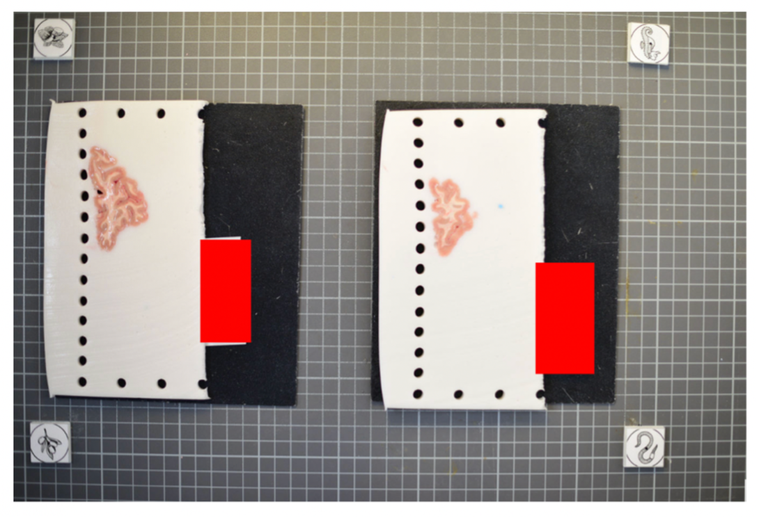
The fiducials form the four corners of the corrected image; hence, all tissue should be within the rectangle formed by the four fiducials. The following image shows slab photos where the fiducials were not placed far enough apart.

Drawing a rectangle through the four fiducial markers makes it clear that much of the tissue lies outside of the pixel-corrected region.

In summary, here is an example of an optimal photographic setup: a uniform, high-contrast background, the provided fiducials located at known distances, and consistent lighting on all slabs.

Tip: It does not matter if the slabs are not placed on a grid, as long as the distance between the fiducials is known. The software will draw digital rulers on your images during pixel correction.
Part 2: Preprocessing Photographs with the Dissection Photo App
The dissection photo app has five principal windows for each subject: Add photos, Correction, Segmentation, Connected components and Reconstruction. The step bar at the top of the window shows which step is being done. Users can backtrack by clicking any of the previous steps at any time, or click the house icon to return to the Projects screen with a summary of all reconstructions done or in process. For each project, work is saved as you go, so the app can be closed at any time and you can pick up later where you left off.
Before continuing, make sure the development version of FreeSurfer is properly sourced, you have downloaded the model weights, and have set NNUNET_MODEL_DIR as an environment variable pointing to the segmentation model. To start the application, use the command dissection_photo_2.
Projects Page
Each specimen corresponds to a project: a folder which will be populated with the outputs from all steps. The project home contains a summary of all projects and their progress.
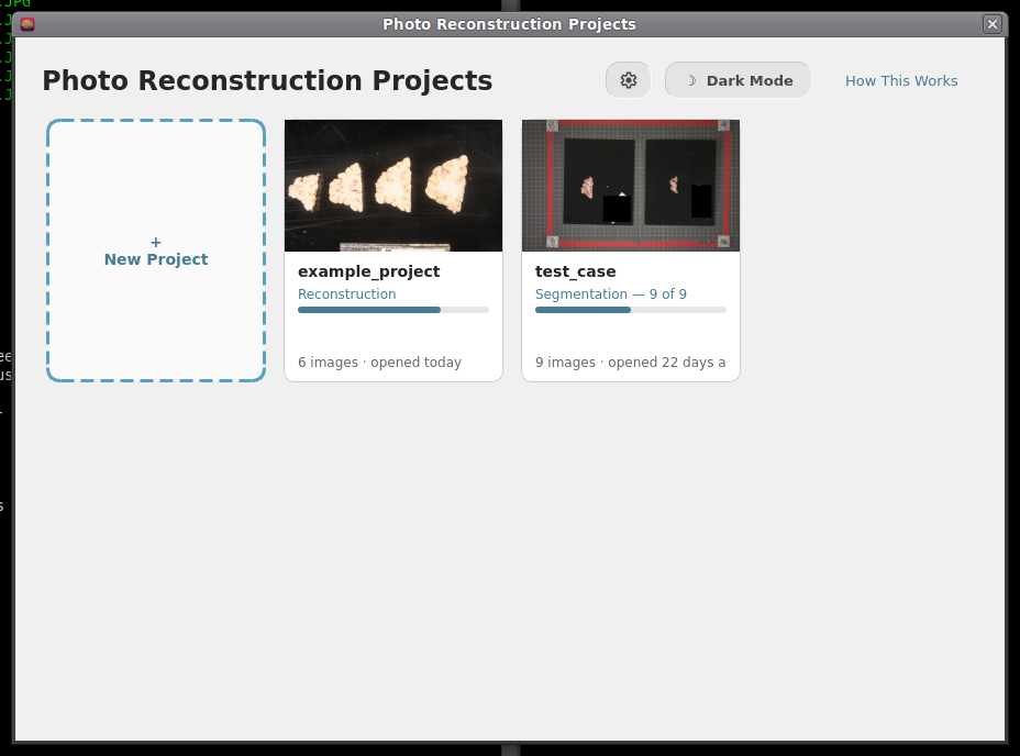
Projects Home screen.
To start processing a new batch of dissection photographs click on New Project.
- Open a project to continue where you left off or backtrack to any completed steps.
Tip: For advanced users, the gear icon can be used to change which resources are used by the automatic segmentation model, covered in automatic segmentation settings.
After naming a project you will be prompted to save a folder of the same name. It is in this folder that the outputs of all steps will be stored.
Directory Structure
For each project the following directory structure will be populated with all outputs.
Folder / file |
Contents |
photos/ |
Copy of the original photographs |
deformed/ |
Pixel-corrected photos |
mask/ |
Binary tissue masks |
connected_components/ |
Label maps per slab |
recon/ |
The 3D-photo reconstruction |
Step 1. Add photographs
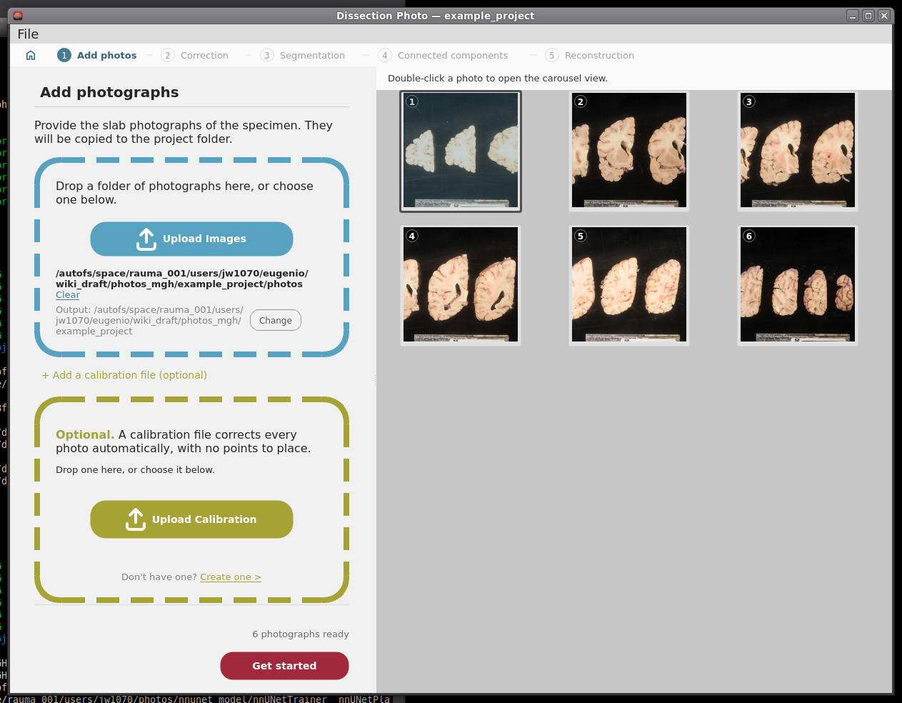
Drag and drop a folder containing dissection photographs, or click Upload Images.
- Select the photographs that should be used for the reconstruction. This can be used to omit repeat photographs, dissection photographs of cerebellar tissue, or gross photographs done prior to slabbing. Double click on any image in the grid for a larger preview.
If you have or would like to create a calibration file to automate pixel correction, click on + Add a calibration file (optional) and follow the steps in Making a Calibration File and then Prospective Correction.
Click Get started.
|
|

Optional: Making a calibration file
This step only needs to be done once, as long as the markers are not moved. For more information on proper usage refer to the Markers for pixel and perspective corrections section in this wiki page.
On the Add photos menu, click + Add a calibration file (optional) to expand the calibration file box, and click on Create one under the Upload Calibration button.
On the next menu, if you have not printed out the fiducial markers, they can be downloaded for printing by clicking the Download Marker Printout button.
- Next, place markers at a known distance in a rectangle formation on the photography table. Be sure that opposite sides of the rectangle created by the fiducial markers are parallel; inaccurate marker placement can lead to incorrect perspective correction.
- Upload the photograph of the empty dissection table with just the markers present, and enter the width and height of the fiducial box.
- For each of the markers, click its centre and outer circle. Options to zoom, pan, and redo/undo can help fix mistakes and make the best calibration possible.
Click Save Calibration and save your calibration file in an easy-to-access location.
Tip: Because of slight lens distortion, the overlaid labels for the outer circle of the markers might not always align throughout the whole circumference. This is not an issue.

The photograph of the empty table with the four markers, uploaded.
|
|


Step 2: Pixel Correction
This step corrects all photographs to be at the same pixel size. Additionally, it can correct for perspective when using a calibration file, or when doing manual correction in 4-point mode.
Prospective Correction (Using fiducial markers and calibration file)
- Every photo is corrected automatically as soon as Correction opens. The markers are found in each photo separately, so it does not matter if the camera moved between photos.
Check each photo: Original shows the photo as taken, with the marker centres that were found; Corrected shows the result. A green tick in the strip at the bottom marks each corrected photo.
If the markers cannot be found in a photo, the app says which ones. On each of them, click the centres of the four markers on the Original view and click Correct this photo.
Retrospective Correction (Using a spatial reference within the photographs)
Choose how many points you can mark, enter the real distances, and place the points on each photo:
Mode |
You need |
Corrects |
2 Point |
One ruler or other known distance (entered as Ruler width (mm)) |
Pixel size only |
3 Point |
Three corners of a rectangle, e.g. two rulers at right angles |
Pixel size, approximate perspective |
4 Point |
Four corners of a rectangle |
Pixel size and perspective |
Ctrl + left click places a point; once all are placed, Ctrl + click moves the nearest one. Clear points starts again.
Click Correct this photo (Ctrl + W also moves on to the next photo). Place points on the Original view; the Corrected view is for checking.
|
|
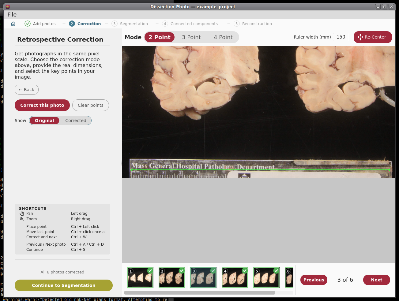
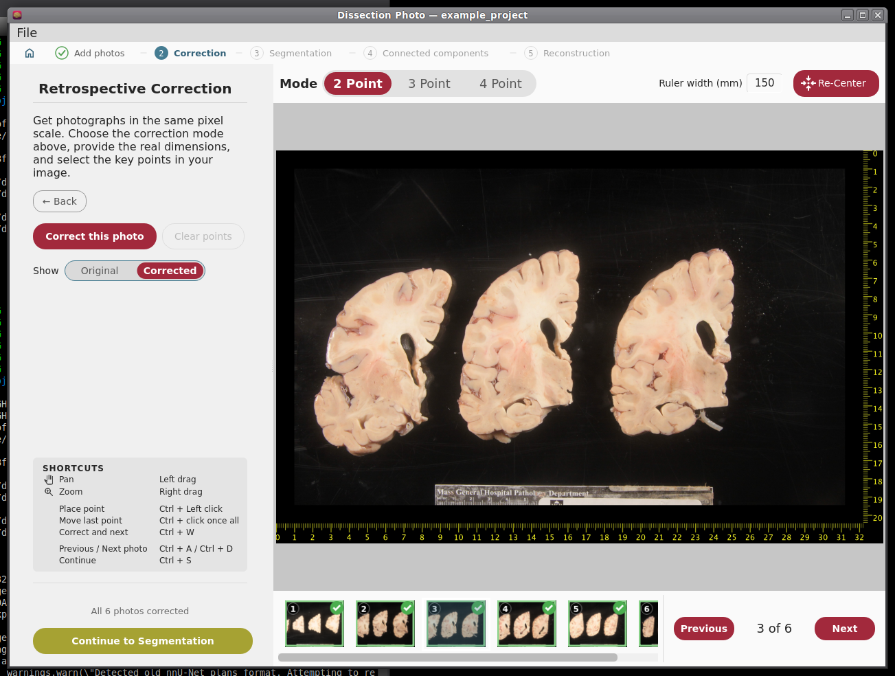
When every photo is corrected, make sure to click Continue to Segmentation.
Step 3: Binary Segmentation
While you were correcting, the app was already finding the tissue in each corrected photo. Here you check each mask and fix it.
|
|
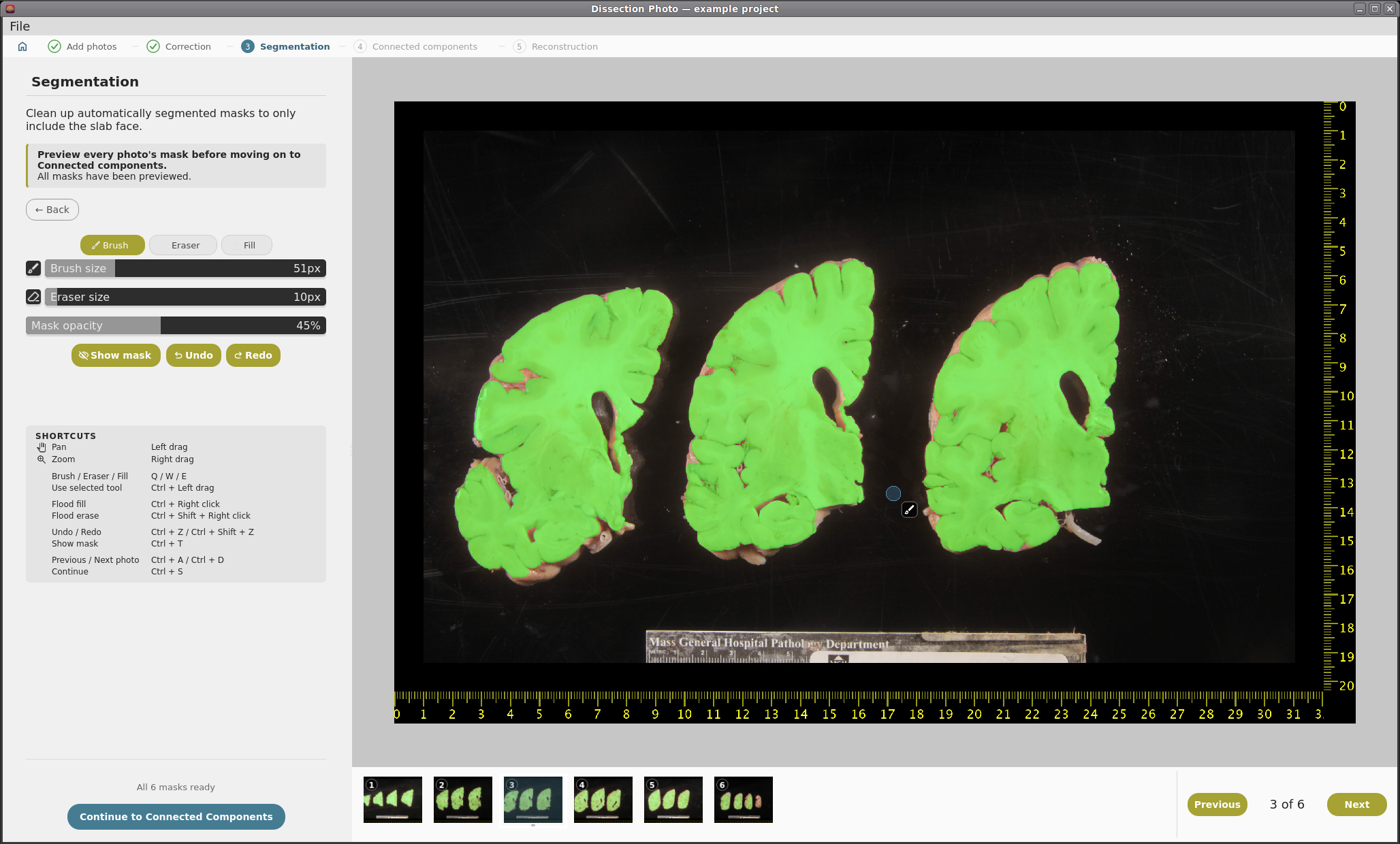
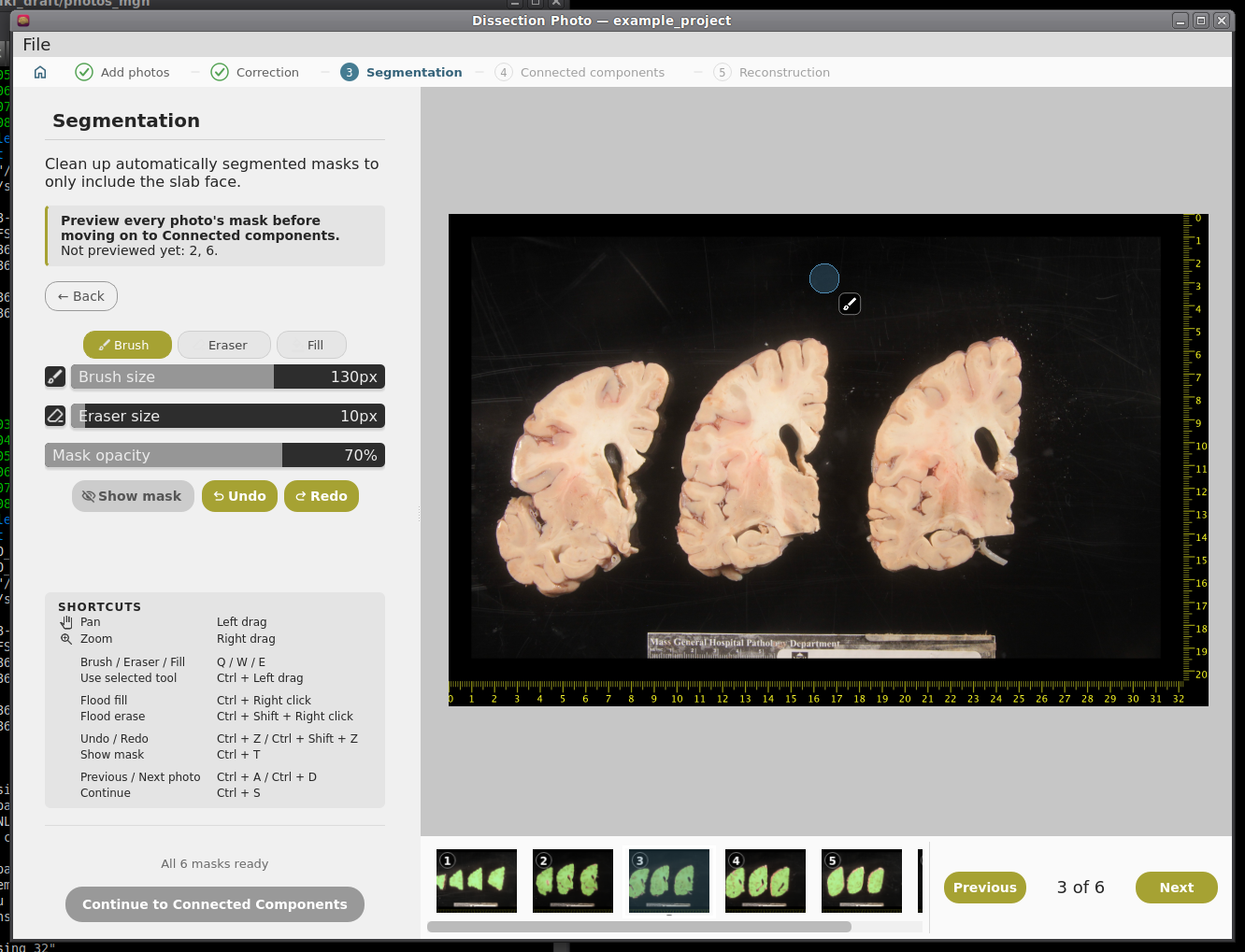
Brush (Q) adds to the mask, Eraser (W) removes from it, and Fill (E) fills an area. Apply the tool with Ctrl + left drag; set the sizes with the sliders.
Mask opacity changes how see-through the mask is; Show mask (Ctrl + T) hides and shows it. Undo and redo with Ctrl + Z and Ctrl + Shift + Z.
- Leave out everything that is not slab tissue: the background, labels, and fragments that do not belong to a slab.
IMPORTANT: the cortical surface should not be part of the masks, so the first slab (if photographed from the anterior side) or the last slab (if photographed from the posterior side) should NOT be included.
You need to look at every photo's mask before Continue to Connected Components becomes available; a note at the top says which ones are left.
Automatic Segmentation Settings
Open them from the gear on the Projects screen. Changes apply the next time photos are segmented.

Automatic segmentation settings.
Setting |
What it does |
Automatic segmentation (On / Off) |
Off: every mask is drawn by hand from scratch. Only turn it off if the automatic segmentation fails on your photos. |
Model folds |
Automatic: 5 folds on a GPU, 1 on the CPU. Five folds are steadier but 4-5x slower. |
Run on |
Automatic uses the GPU if there is one; or choose GPU or CPU. |
Maximum threads |
The most processor threads segmentation may use. If the computer has fewer, it uses all it has. |
Model folder |
The folder of the segmentation model (nnUNetTrainer__nnUNetPlans__2d). |
Step 4: Slab Connected Components
Here you tell the app which pieces of tissue belong to which slab.
- In each photo, draw a box inside each slab (Ctrl + left drag), always in the same order: anterior to posterior, or posterior to anterior. The slab is colored as soon as its box is drawn.
- Keep boxes small and inside the tissue, and do not let them overlap. If a slab is in several pieces (e.g. a detached temporal lobe), make sure its box touches every piece.
Click Save components (Ctrl + W also moves on to the next photo); moving to another photo saves it too. A green tick marks saved photos. If you change a saved photo, its tick goes away until you save it again.
Undo and redo with Ctrl + Z and Ctrl + Shift + Z. Slab overlay changes how strongly the slabs are colored.
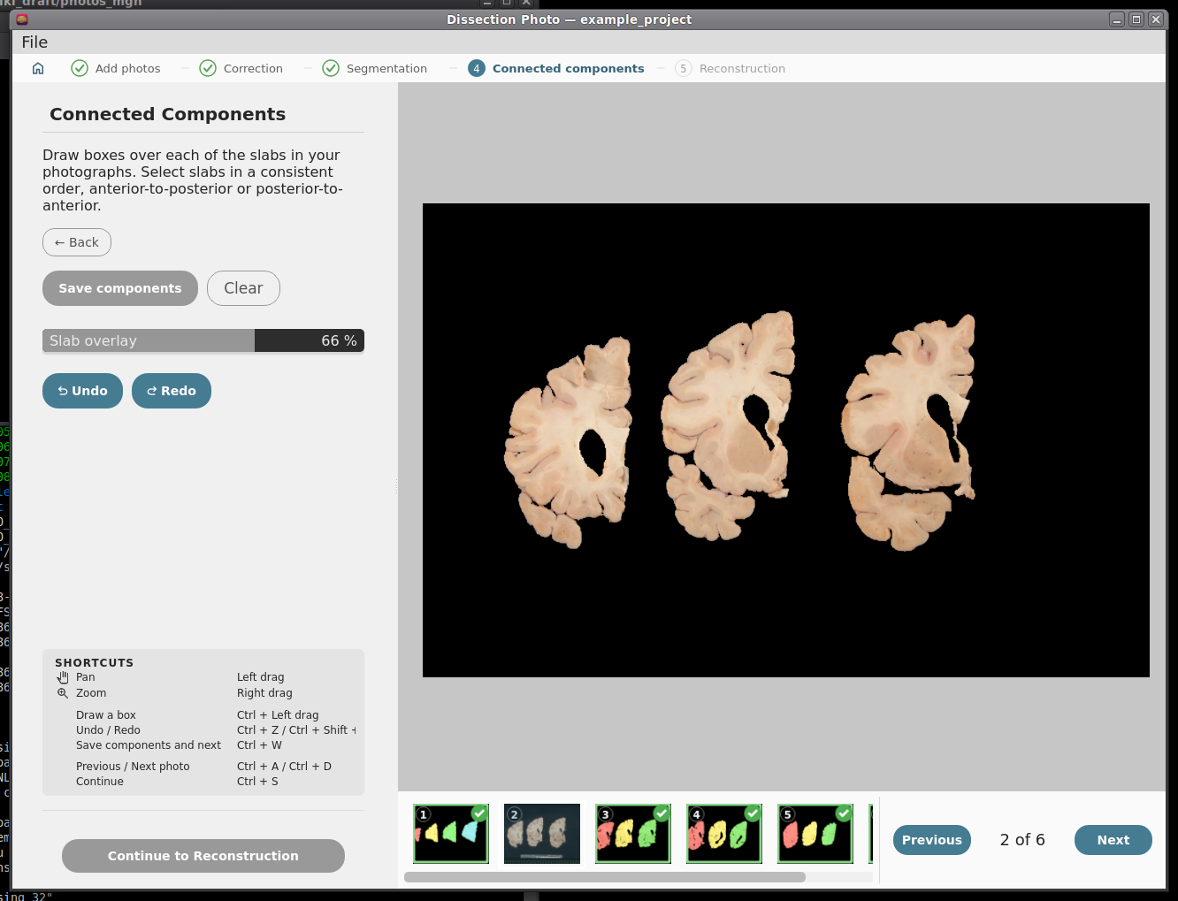
Before any boxes are drawn.
|
|
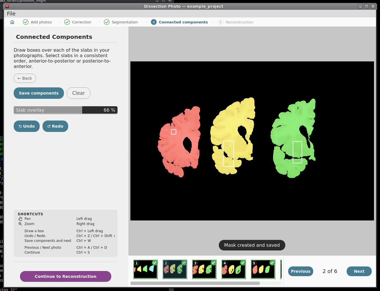
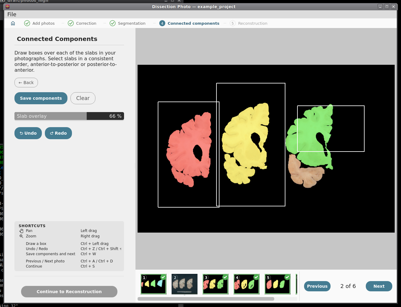
Step 5: Photo Reconstruction
Please see our separate page on the command mri_3d_photo_recon. The last step runs mri_3d_photo_recon on the images of the project. While the reconstruction is running in the background the user can continue processing images of other cases.
Output folder: the folder inside the project for the results (default recon).
Slabs: the hemisphere, the slab thickness in mm, which face of the slabs was photographed (anterior or posterior), the order the photos were taken in, and whether the tissue is fixed or fresh.
Reference: none (the MNI atlas), a surface mesh (.stl), an MRI scan, or both. See Which reference should I use?
- Optionally: the number of threads, a graphics card, and the advanced settings. Empty expert weights use the command's own defaults, shown in grey.
Click Run Reconstruction.
The command being run is shown on the right (Copy command copies it, for example to run it on another computer), and its output appears in the log below it. The reconstruction keeps running if you switch to another project; the Projects screen shows its progress.

A reconstruction running, with the command and its log.
Part 3: Downstream Tasks
Photo-Synthseg Segmentation
The 3D reconstructed stacks can be segmented with SynthSeg using the flag --photo. Please see the wiki page for SynthSeg for further information.
|
|
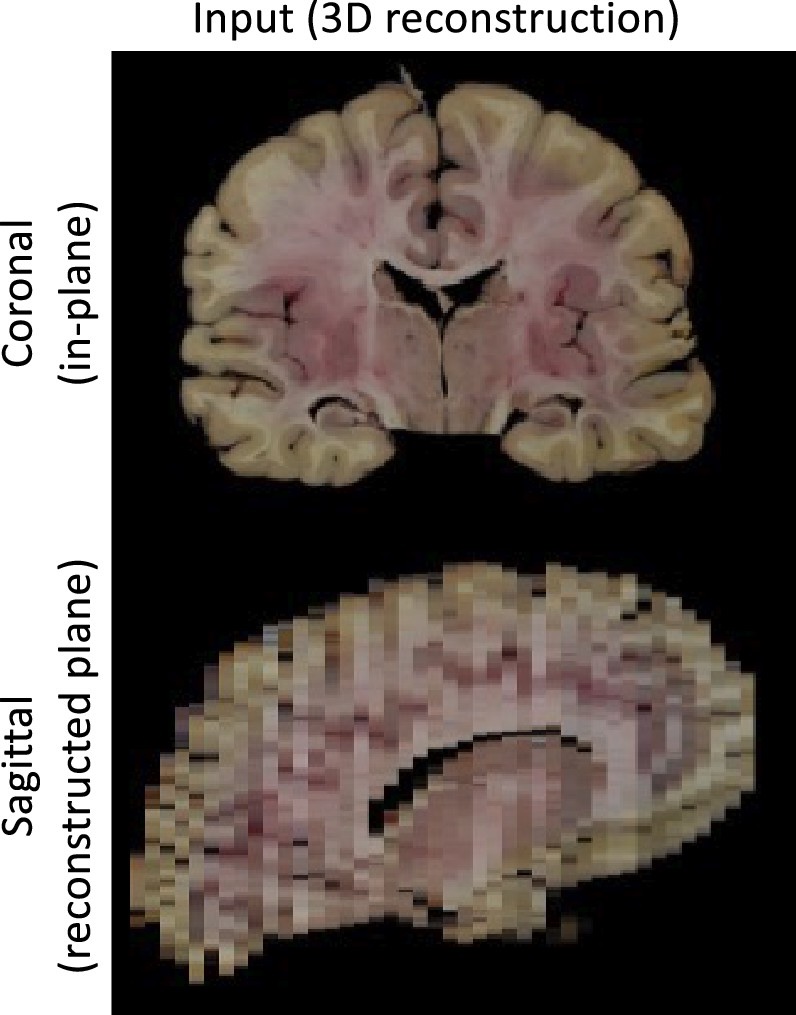

Example for running SynthSeg on a Left Hemisphere
mri_synthseg \
--i ./recon/photo_recon.mgz \
--o ./recon/photo_recon_synthseg.mgz \
--photo left \
--parc